
 Glaucoma Diagnosis & Treatment
Glaucoma Can Lead to Blindness or Severe Vision Loss if not Treated Timely and Properly
What is Glaucoma?
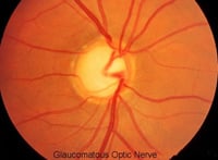
Glaucoma is a group of diseases that can damage the eye's optic nerve and result in vision loss and blindness. The exact location of the disease damage is in the retinal nerve fiber layer, one of the ten layers of the retina. There is a characteristic acquired loss and/or degradation of retinal ganglion cells (retinal nerve fiber layer).
The optic nerve is a bundle of more than 1 million nerve fibers. It connects the retina to the brain. A healthy optic nerve is necessary for good vision.
Glaucoma is a multifactorial, progressive, neurodegenerative disorder. Glaucoma is characterized by the death of retina ganglion cells, optic nerve atrophy and loss of neurons in the brain (lateral geniculate nucleus and the visual cortex). In primary open-angle glaucoma, intraocular pressure (IOP) is the biggest risk factor for glaucoma onset and progression and is the only risk factor glaucoma specialists can modify. However, neurological factors contribute to glaucoma's onset and progression that unfortunately cannot be modified. There is ongoing research to develop treatment to minimize the neurological damage for glaucoma patients.
Facts About Glaucoma
- Approximately three million Americans have glaucoma. However, almost half of these people are unaware that they have the disease.
- 800,000 Americans are legally blind from glaucoma.
- Glaucoma causes a loss of some of the nerve fiber layer in the retina of the eye.
- There is no cure for glaucoma. In most cases, glaucoma can however be treated with medicine (eye drops) to prevent or reduce further vision loss.
- Once vision is lost it cannot be recovered. Therefore, it is imperative to prevent loss by diagnosing and treating glaucoma early.
- The following are risk factors for glaucoma:
- High intraocular pressure (aqueous fluid pressure inside the eye)
- Family history of glaucoma
- Over age of 40
- Hispanic
- African-American
- Diabetes
- High Myopia (nearsightedness)
- Previous eye injury
- Corneal thickness less than average
In the front of the eye is a space called the anterior chamber. A clear fluid flows continuously in and out of the chamber and nourishes nearby tissues. The fluid leaves the chamber at the open angle where the cornea and iris meet. When the fluid reaches the angle, it flows through a spongy meshwork, like a drain, and leaves the eye.
Sometimes, when the fluid reaches the angle, it passes too slowly through the meshwork drain. As the fluid builds up, the pressure inside the eye rises to a level that may damage the optic nerve. When the optic nerve is damaged from increased pressure, open-angle glaucoma and vision loss may result. That's why controlling pressure inside the eye is important. However, increased eye pressure does not mean you have the disease, but it does mean you are at risk for glaucoma. A person has glaucoma only if the optic nerve or retinal nerve tissue is damaged.
Not every person with increased eye pressure will develop glaucoma. Some eyes can tolerate higher internal eye pressure better than others. Whether you develop glaucoma depends on the level of pressure your optic nerve can tolerate without being damaged. This level is different for each person. That's why a comprehensive dilated eye exam and especially a Spectral Domain OCT scan (optical coherence tomography) test is very important. In contrast, glaucoma can develop without increased eye pressure. This form of glaucoma is called low-tension or normal-tension glaucoma.
At first, open-angle glaucoma has no symptoms. It causes no pain and vision stays normal. As glaucoma remains untreated, people may miss objects to the side and out of the corner of their eye. Without treatment, people with glaucoma will slowly lose their peripheral (side) vision and some areas within the central visual field. Over time, straight-ahead vision and peripheral vision may decrease until no vision remains.
Glaucoma Signs and Symptoms
THERE ARE NONE! In the most common form of glaucoma there are no signs and symptoms obvious to the patient until very late stages of the disease. It is then too late!
Have your eyes examined every year by a glaucoma specialist or eye doctor!
Glaucoma Diagnostic Tests
Glaucoma is detected through a series of special tests as well as a general comprehensive eye exam that includes:
Optical Coherence Tomography (OCT Scan)
Optical Coherence Tomography (OCT Scan) Can Diagnose Glaucoma Before All Other Tests. It is arguably the most accurate test in the world to assist our doctors in diagnosing glaucoma.
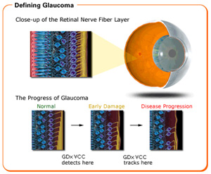
GDxVCC Scanning Laser Polarimetry
GDxVCC Scanning Laser Polarimetry: This is also one of the world’s most accurate single tests in diagnosing glaucoma. Along with the OCT scan, this is the only type of technology available that directly and objectively evaluates the retinal nerve fiber layer of the eye, which is the specific site of damage from glaucoma. OCT and GDxVCC can detect nerve fiber layer loss approximately six years before visual field loss.
Scanning Laser Polarimetry or an OCT scan is a must in diagnosing and monitoring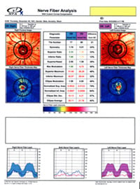 glaucoma. The newest spectral domain OCT used by Master Eye Associates' optometric glaucoma specialists provides even better diagnostic capabilities than the GDx or the Stratus OCT because our new OCT can monitor progression of retinal nerve fiber loss over time. Only the newest spectral domain OCT can monitor progressive loss of the nerve fiber layer. That function is crucial in monitoring glaucoma, which by its very nature is a disease of progression.
At Master Eye Associates we believe that having the best technology is a way of showing that we care about our patients.
Visual acuity test. This eye chart test measures how well you see at various distances.
Visual field test. This test measures your central and peripheral vision  area. It helps tell if you have reduced visual sensitivity to certain areas within your visual field as well as lost visual field areas called scotomas. Although this test is useful and still the most used test in diagnosing glaucoma it is not as accurate in diagnosing early glaucoma as the GDxVCC. Unfortunately, up to 60 percent of nerve fibers may be diminished or damaged before visual field analysis can identify a problem.
Remember, the OCT scan or our GDxVCC threshold exam can identify glaucoma up to six years before the problem is identified on the visual field test.
Dilated eye exam. Drops are placed in your eyes to widen, or dilate, the pupils. Your eye care professional uses a special magnifying lens to examine your retina and optic nerve for signs of damage and other eye problems. After the exam, your close-up vision may remain blurred for  several hours.
Tonometry. An instrument called a tonometer measures the pressure inside the eye. There are several different methods to test the fluid pressure inside the eyes. The newest tonometer takes into account the many biomechanical properties of the cornea that influence intraocular pressure.
Pachymetry. A numbing drop is applied to your eye. Your eye care professional uses an ultrasonic wave instrument to measure the thickness of your cornea. It is now well documented by a major national study (Ocular Hypertension and Treatment Study OHTS) that thinner than average corneas are a definite risk factor for glaucoma. This test can also be performed using optical coherence tomography instead of ultrasound.
Glaucoma Treatments
At this time there is no cure for glaucoma. Vision lost from the disease cannot be restored. That is why immediate treatment for early stage, open-angle glaucoma is so important. Immediate treatment can delay progression of the disease. Glaucoma treatments include eye drop medicines, laser trabeculoplasty, conventional surgery, or a combination of any of these. While these treatments may save remaining vision, they do not improve sight already lost from glaucoma.
Glaucoma Almost Always Gets Worse
Glaucoma is a progressive disease and much of the treatment is based on the rate of progression of the disease. Unfortunately, no matter how well the disease is treated, no matter how good the glaucoma specialist, no matter how good the medicine, glaucoma usually gets worse. The goal is to maintain functional vision for the patient's lifetime.
Treatment with Medicine/Eye Drops
Medicines, in the form of eye drops or pills, are the most common early treatment for glaucoma. Some medicines cause the eye to make less fluid. Others lower pressure by helping fluid drain from the eye. Glaucoma medicines are often instilled several times a day. Because glaucoma has no symptoms, people may be tempted to stop taking, or may forget to take, their medicine. You need to use the drops or pills as long as they help control your eye pressure. Regular use is very important. Make sure your eye care professional shows you how to put the drops into your eye.
Most people have no problems. However, some medicines can cause headaches or other side effects. For example, drops may cause stinging, burning, and redness in the eyes. If you have problems with one medicine, tell your eye care professional. Treatment with a different dose or a new drug may be possible. It is also important that you tell your eye care professional about other medicines you may be taking before you begin glaucoma treatment. Sometimes the drops can interfere with the way other medicines work.
Click for a list of currently used glaucoma medicines and their side effects and benefits.
Glaucoma Surgeries
If glaucoma eye drop medicine does not adequately control the intraocular pressure or the damage to the eye from glaucoma, glaucoma surgery will be recommended. There are many options in glaucoma surgery such as:
Argon Laser Trabeculoplasty (ALT)
Selective Laser Trabeculoplasty (SLT)
Normal - Low Tension Glaucoma
In low-tension or normal-tension glaucoma, optic nerve damage and narrowed side vision occur in people with normal eye pressure. Lowering eye pressure at least 30 percent through medicines slows the disease in some people. Glaucoma may worsen in others despite low pressures. A comprehensive medical history by a glaucoma specialist is important in identifying other potential risk factors, such as low blood pressure, that may contribute to low-tension glaucoma especially while sleeping. If no risk factors are identified, the treatment options for low-tension glaucoma are the same as for open-angle glaucoma.
Angle-closure Glaucoma
In angle-closure glaucoma, the fluid at the front of the eye cannot reach the angle and leave the eye. The angle gets blocked by part of the iris. People with this type of glaucoma have a sudden increase in eye pressure. Symptoms include severe pain and nausea, as well as redness of the eye and blurred vision. If you have these symptoms, you need to seek treatment immediately. This is a medical emergency. If your doctor is unavailable, go to the nearest hospital or clinic. Without treatment to improve the flow of fluid, the eye can become blind in as few as one or two days. Usually, prompt laser surgery and medicines can clear the blockage and protect sight.
Vision Loss from Glaucoma
If you have lost some sight from glaucoma, ask your eye care professional about low vision services and devices that may help you make the most of your remaining vision. Ask for a referral to a specialist in low vision. Many community organizations and agencies offer information about low vision counseling, training, and other special services for people with visual impairments.
You also can help protect the vision of family members and friends who may be at high risk for glaucoma--African Americans over age 40; everyone over age 60, especially Hispanic Americans; and people with a family history of the disease. Encourage them to have a comprehensive dilated eye exam at least once every two years.
Remember the key to maintaining your eyesight with glaucoma is:
-
To diagnose it at the earliest possible time
-
Lowering eye pressure in glaucoma's early stages slows progression of the disease and helps save vision.
