
OCT Scans are Used to More Accurately Diagnose & Treat Retinal Eye Diseases and Glaucoma
Optical Coherence Tomography provides better resolution than an MRI and Helps Diagnose Retina & Corneal Disease and Glaucoma
Optical Coherence Tomography (“ OCT”) is a painless, non-contact, non-invasive imaging technique used to obtain high resolution cross-sectional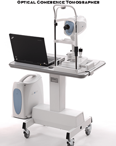 and three dimensional images of the retina, cornea and anterior chamber of the eye. OCT provides images at much higher clarity and resolution than other imaging instruments such as MRI or ultrasound. Optical Coherence Tomography obtains sub-surface images of translucent or opaque materials at a resolution equivalent to a low-power microscope.
How OCT Works
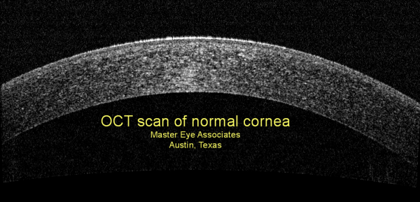
OCT produces cross-sectional images and three-dimensional imaging of the cornea and the anterior segment of the eye and the retina using the optical backscattering of light similar to B-scan ultrasonography. It images reflections from within tissue to provide cross-sectional views. The physics principle allowing the filtering of scattered light is optical coherence. OCT delivers high resolution because it is based on light, rather than sound or radio frequency. An optical beam of infrared light is directed at the tissue and a small portion of this light that reflects from sub-surface features is collected. Most of the light is scattered instead of reflected.
 OCT and ultrasound use echo location principles which rely on sound or light to navigate and locate. Other medical imaging techniques such as computerized axial tomography (CAT scan), magnetic resonance imaging (MRI), or positron emission tomography (PET scan) do not utilize the echolocation principle.
The OCT technique is limited to imaging 1 to 2 mm below the surface in biological tissue because at greater depths the proportion of light that escapes without scattering is too small to be detected or measured. No preparation of the human tissue is required and OCT scans are safe using near infrared light. Using long wavelength light allows it to enter the biological tissue, which is the scattering medium of the light.
Leading Edge Technology & Instruments are Another Way of Showing that We Care About our Patients
Master Eye Associates has an ultra-high speed, high resolution Optical Coherence Tomographer (“OCT”) retina scanner used for retina imaging, corneal scans, anterior chamber views and glaucoma analysis and diagnosis. Our OCT instruments use the newest generation Fourier-Domain Optical Coherence technology that has just recently emerged from clinical research. Our new OCT instruments feature ultra-high speed and high resolution to visualize the retinal tissue with ultra-high clarity in a fraction of seconds.
 The OCT instruments used by Master Eye Associates also images and scans both the anterior and posterior part of the eye. Some other OCT instruments cannot image the anterior part of the eye, which is crucial in diagnosis of many eye diseases.
Fourier Domain OCT vs. Time Domain OCT
Master Eye Associates utilizes the new, greatly improved OCT instrument. The old generation OCT is based on the Time Domain OCT technology which performs very slow retinal scans and limits its ability to provide scans as accurately and as clearly as the new OCT technology. Our new OCT technology is 6500% faster than the Stratus OCT, which uses Time Domain technology. This provides much greater benefits and accuracy for our patients. Our OCT also provides 200% greater clarity and resolution than the most common used technology.
Additionally, the new Fourier Domain OCT is able to provide an analysis of progression of the disease for glaucoma scans. Glaucoma is a disease of progression and the ability to monitor progression is vital. Only the new OCT can perform this function. The older OCT’s cannot track progression.
Master Eye Associates is always on the leading edge of ocular imaging. We can analyze or perform the following:
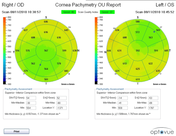
- Mapping of corneal thickness
- Keratoconus corneal evaluation
- Measurement of LASIK flap and stromal bed thickness
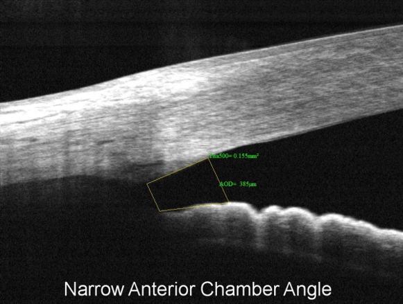
- Visualization and measurement of anterior chamber angle and diagnosis of narrow angle glaucoma
- Measuring the dimensions of the anterior chamber and assessing the fit of intraocular lens implants
- Visualizing and measuring the results of corneal implants and lamellar procedures
- Imaging through corneal opacity to see internal eye structures
- Posterior Vitreous detachment
- Macular Holes
- Epiretinal membrane
- Macular Degeneration
- Abnormal fluid accumulation within the retina
- Central serous retinopathy
- Retinal pigment epithelial detachment
- Cystoid macular edema
- Retinoschisis
- Diabetic Retinopathy
- Retinal detachments
- Optic nerve diseases
- Optic nerve edema
- Congenital pit of the optic nerve
- Choroidal nevus


Master Eye Associates – Experienced Eye Care You Can Trust!
